UNDERSTANDING EDEMA: MEANING, FORMATION, CAUSES AND TISSUE FLUID FORMATION.
INTRODUCTION
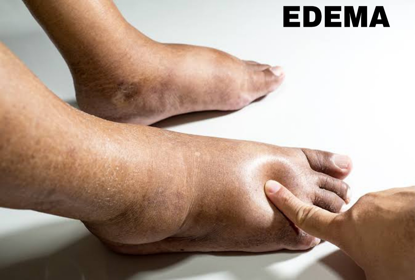
Edema is the swelling that occurs when your body traps excess fluid especially in the skin. This fluid is known as tissue fluid or interstitial fluid (ISF).
The interstitial fluid is defined as the fluid which fills the spaces between cells. It helps to exchange materials between cells, between blood (plasma) and tissue cells and waste removal.
PLASMA IS THE LIQUID PART OF THE BLOOD. THE SOLID PART OF THE BLOOD IS COMPOSED OF CELLS.


The interstitial fluid and plasma are kept in a physiological balance state to maintain their functions but in some disease conditions this balance is altered and if positive on the interstitial fluid, it results to edema.
NOTABLE CAUSES INCLUDE:
Allergy, inflammation, trauma, sitting or standing for a long time, medications, starvation, malnutrition, pregnancy, organ failures, vascular obstructions, poor drainage of a body area are known to cause edema.
FLUID EXCHANGE
The part of your blood vessels responsible for fluid exchange with the interstitial space (space containing interstitial fluid) are the arterioles (arterial ends) and venules (venous ends).
Arteries take blood from your heart to other organs while veins bring the blood back to your heart.
FORCES GOVERNING FLUID EXCHANGE BETWEEN THE BLOOD VESSELS (CAPILLARIES AND VENULES) AND INTERSTITIAL SPACE.
1- Hydrostatic pressure
2- Oncotic pressure/ colloid osmotic pressure
HYDROSTATIC PRESSURE
This is the driving force for filtration of water and other substances from blood to tissue spaces. It is about 30 to 37mmHg at arterial ends and 15 to 17mmHg at venous ends.
ONCOTIC PRESSURE
This is the pressure exerted by large molecules in your plasma called plasma proteins. These molecules are large enough that in a normal condition they can't cross your vessel wall and enter the interstitial space.
Their function is to draw filterd fluid back into the blood vessel. So as hydrostatic pressure filters fluid out the vessel, oncotic pressure draws this fluid back into the vessel.
The oncotic pressure is about 25mmHg through the circulation. IT WORKS AGAINST EDEMA FORMATION.
FORMATION OF TISSUE FLUID
This involved two processes:
1- Filtration
2- Reabsorption
FILTRATION
Your tissue fluid is formed by filtration. As hydrostatic pressure at arterial end (30mmHg) is greater than oncotic pressure (25mmHg), fluid is filtered.
REABSORPTION
Filterd fluid at arterial end is reabsorbed back through venous end because oncotic pressure (25mmHg) is greater than hydrostatic pressure at venous end (15 to 17mmHg).


From the pictures above, the lymph capillary helps to drain fluids and waste from the interstitial space, thereby striving to maintaining balance of the fluids too. Lymph is the fluid containing wastes, toxins and infection-fighting white blood cells.
TYPES/CLASSIFICATION OF EDEMA
1- INTRACELLULAR EDEMA
2- EXTRACELLULAR EDEMA
INTRACELLULAR EDEMA
The cells on their own can swell too when water accumulates in them. Sodium is a water-loving element and your cells possess a pump( sodium-potassium ATPase pump).that pumps sodium out of them. When the pumping activity of this pump is altered, sodium accumulates in the cells, drawing water with it into the cells cause swelling of the cell.
This type of edema is caused by malnutrition, inflammation, poor metabolism, poor blood flow to the cells.


EXTRACELLULAR EDEMA
Occurs from conditions that make your interstitial fluid pressure to be positive. Example are heart failure, renal disease and lymphatic obstruction.


Edema can also be classified as:
1- PITTING EDEMA
2- NON-PITTING EDEMA
PITTING EDEMA
Here, when a pressure is applied by a finger on the swelling, it leaves a depression (pit) which disappears about 30 seconds when the pressure is removed. The depression is due to disappearance of fluids to other areas which later flows back after removal of the pressure.

NON-PITTING EDEMA
Due to coagulation of the fluids causing the swelling, depression do not occur when a pressure is applied. That is fibrosis has occurred. Such is seen in the case of elephantiasis or lymphoedema.

Scrotal lymphoedema


In elephantiasis, there is lymphatic obstruction by a warm know as filaria warm.
CAUSES OF EDEMA:
1- FACTORS THAT WILL INCREASE CAPILLARY HYDROSTATIC PRESSURE
A-- INCREASE IN VENOUS PRESSURE:
Caused by obstruction by clots, right heart failure.
B-- ARTERIOLAR DILATION:
Seen in hot weather, standing or sitting for a long time. Standing for a long time can cause edema in the legs.
C-- SALT AND WATER RETENTION:
This causes increase in fluids outside your cells resulting in increased tissue fluid.
D-- KIDNEY DISEASE ( KIDNEY FAILURE)
Here the kidney does not excrete urine as it should when functions are normal. There's build up of fluid resulting in edema.
2- FACTORS THAT DECREASE ONCOTIC PRESSURE
A-- MALNUTRITION
These plasma proteins responsible for oncotic pressure is made from the food we eat. Malnutrition doesn't favour the production of these molecules as there's no nutrient for that.
B-- KIDNEY DISEASES (NEPHROSIS, NEPHROTIC SYNDROME)
Here, there's large amount of protein leakage into the urine thereby decreasing oncotic pressure and causing edema.
C-- INCREASE CAPILLARY PERMEABILITY
Occurs in inflammation, burns etc. Proteins leak into interstitial space thus reducing oncotic pressure in the arterioles.
3- INADEQUATE TISSUE DRAINAGE
A-- OBSTRUCTION OF LYMPHATICS
This obstruction can be by parasites
B-- SURGICAL REMOVAL OF LYMPH NODES
Seen in breast cancer treatment, there's a problem with lymphatic drainage of the arm.
IN PREGNANCY, EDEMA RESULTS FROM INCREASE IN BODY FLUID AND PRESSURE OF THE FOETUS ON THE PELVIC REGION. THIS PRESSURE COMPRESSES THE VAINS DRAINING THE LEG CAUSING FLUID BUILD UP.


Comments
Post a Comment